
Beyond the Diagnosis: Why Clinical Precision Starts with Defining the Brain Injury Mechanism
For any rehabilitation professional, encountering a client with an acquired brain injury represents one of the most complex and multi-faceted challenges in clinical practice. Because the brain dictates nearly every aspect of human existence, a single injury can disrupt a client’s movement, sensation, memory, emotional regulation, and vital autonomic functions simultaneously.
When junior practitioners or students transition into neuro-rehabilitation, it is easy to become overwhelmed by the sheer volume of symptoms presenting at once. However, effective therapeutic planning cannot begin with a reactive approach to symptoms. Long-term clinical success requires us to pause, look past the surface-level diagnosis, and establish a rigorous baseline understanding of the exact mechanism of the injury.
Before we can design an intervention to reshape neural pathways, we must completely understand how those pathways were disrupted.
Classifying the Genesis: Traumatic vs. Non-Traumatic Causes
The umbrella term Acquired Brain Injury (ABI) encompasses any damage to the brain that occurs after birth. To build an accurate clinical framework, a practitioner must first differentiate between the two primary pathways of injury:
- Traumatic Brain Injuries (TBI): Resulting from an external physical force, such as a fall, motor vehicle collision, or assault, which causes structural damage through acceleration, deceleration, or penetration.
- Non-Traumatic Brain Injuries: Resulting from internal cellular stress, such as a lack of oxygen (anoxia or hypoxia), metabolic crises, toxic exposure, or stroke.
Understanding this initial distinction is critical because the underlying pathology dictates how the injury will unfold over time. A traumatic impact introduces unique biomechanical forces, whereas an anoxic event creates widespread metabolic deprivation. Recognizing the source allows clinicians to accurately interpret early clinical data and anticipate potential complications.
The Pathological Cascade: Primary vs. Secondary Mechanisms
A brain injury is not a single, isolated event frozen in time. Instead, it consists of a dual-phase pathological cascade that practitioners must carefully monitor:
- The Primary Mechanism: This is the immediate physical or metabolic damage occurring at the exact moment of insult. Examples include the direct tearing of axons during a traumatic impact or the immediate cellular death caused by a sudden stroke.
- The Secondary Mechanism: This is the subsequent wave of biological destruction that unfolds hours, days, or weeks following the initial injury. It includes neuroinflammation, brain swelling (edema), intracranial pressure spikes, and chemical imbalances that cause further tissue damage.
In a rehabilitation context, understanding secondary mechanisms is essential. While we cannot alter the primary injury that has already occurred, our early therapeutic environments and handling strategies can significantly influence how secondary tissue damage settles, directly affecting the client’s long-term functional recovery.
Mapping the Destruction: Focal vs. Diffuse Injury Types
The spatial distribution of a brain injury fundamentally dictates how symptoms manifest in everyday life. Clinicians must be highly proficient in distinguishing between focal and diffuse pathologies.
Focal injuries are localized to a specific geographic region of the brain. A classic example is an intracranial hemorrhage, where bleeding occurs in a confined space, causing localized pressure and tissue compression. The resulting deficits are typically tied directly to the functional responsibility of that specific area, such as localized motor weakness or a distinct speech impairment.
Conversely, diffuse injuries involve widespread damage scattered across multiple networks. Diffuse Axonal Injury (DAI), frequently caused by rapid rotational or shearing forces during an accident, is a prime example. DAI involves microscopic tearing of the brain’s white matter communication lines. Because diffuse injuries damage the systemic highways connecting different regions, they often manifest as complex, generalized impairments in processing speed, executive functioning, and emotional regulation. These deficits can be incredibly challenging to isolate on standard neuroimaging but have massive implications for everyday functional outcomes.
Translating Metrics to Real-World Functional Outcomes
To communicate effectively within an interdisciplinary medical team, rehabilitation professionals must know how to interpret objective severity classifications and map them to real-world expectations.
Metrics such as the Glasgow Coma Scale (GCS), the duration of a client’s loss of consciousness (LOC), and the length of post-traumatic amnesia (PTA) are not just numbers on an intake form. They are vital predictive markers. By mastering how these acute indicators relate to long-term functional outcomes, a clinician can establish realistic prognoses, manage family expectations, and baseline their therapeutic goals with absolute precision.
Establishing the Indispensable Architecture for Therapy
It is a common pitfall in rehabilitation to rush directly into intervention strategies. However, treating cognitive or physical deficits without a deep understanding of the structural injury is akin to building a house without a blueprint. Groundwork must precede planning.
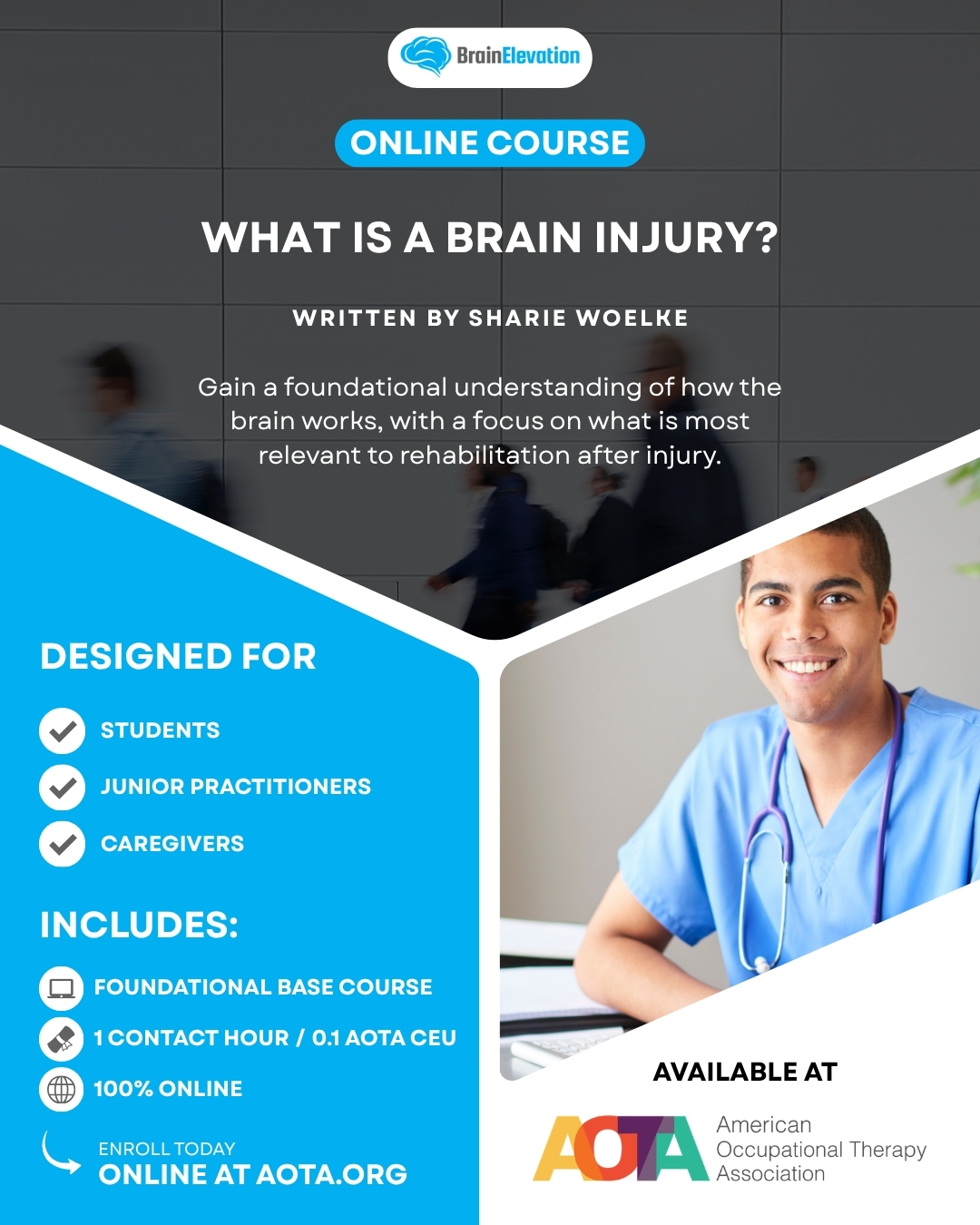
To bridge this specific educational gap, Brain Elevation highlights the foundational course: What is a Brain Injury.
Authored by Sharie Woelke, OT Reg. (Ont), RP, BMR, MEd, Clinical Director of Renew Neurotherapy and Director of Instructional Design for Brain Elevation Consulting, this course is meticulously designed to provide the baseline architecture every practitioner requires. This specialized program focuses entirely on deep clinical comprehension, preparing students, junior practitioners, and clinical caregivers for the advanced therapeutic planning and rehabilitation strategies developed later in the certificate program.
Course Details at a Glance:
- Educational Credit: .1 AOTA CEU / 1 Contact Hour / 1.25 NBCOT PDUs.
- Curriculum Integration: A core component of the Professional Certificate in Applied Brain Injury Rehabilitation and the Brain Basics for Rehabilitation Practice and Recovery Microcredential.
- Key Competencies: ABI definition, primary and secondary tissue damage mechanisms, focal vs. diffuse analysis (including DAI), and severity metric interpretation.
Equip your practice with the scientific integrity and clinical authority your clients deserve. Secure your baseline understanding by enrolling directly through the American Occupational Therapy Association (AOTA).
Enroll in “What is a Brain Injury” on AOTA Here