

Decoding the Injured Brain: Why Network-Level Mastery is Essential for Effective ABI Rehab
For students, junior practitioners, and clinical caregivers stepping into the field of neuro-rehabilitation, the initial learning curve can feel incredibly steep. When an individual sustains an Acquired Brain Injury (ABI), the resulting clinical picture rarely presents as an isolated, predictable set of symptoms. Instead, clinicians are often faced with a shifting matrix of cognitive, emotional, and physical deficits that refuse to fit neatly into basic anatomical text boxes.
The reason for this complexity is fundamental: the brain is the most complex and dynamic organ in the human body. It regulates movement, sensation, memory, emotion, and vital functions all through an intricate web of regions and networks that constantly interact.
To truly support patient recovery, a baseline understanding of basic anatomy is no longer sufficient. Effective neuro-rehabilitation demands mastery at both the microscopic cellular level and the macroscopic network level. Without this dual framework, it is impossible to accurately isolate why specific symptoms emerge or to strategically leverage neuroplasticity for targeted recovery.
The Cellular Reality: Moving Beyond Neurons
When studying brain injury, traditional education frequently over-indexes on the role of the neuron. While neuronal communication is undeniably crucial, true clinical mastery requires an understanding of the entire cellular environment, specifically the vital relationship between neurons and glial cells.
Glial cells are not passive bystanders; they serve critical roles in structural support, metabolic regulation, and immunological defense within the central nervous system. When an acquired brain injury occurs, the disruption is widespread:
- Neuro-trauma immediately compromises the delicate protective structures of the brain, including the meninges, cerebrospinal fluid, and the ventricular system, altering their capacity for injury prevention and increasing vulnerability.
- At the cellular level, injury disrupts the critical communication and regulatory balance between neurons and glial cells.
- This cascade of cellular distress directly contributes to metabolic crises, neuroinflammation, and secondary cellular death, which manifest as the acute symptoms observed clinic-wide.
For junior practitioners and students, learning to differentiate between neuronal and glial disruption provides a clearer clinical picture of what is happening beneath the surface during the early stages of a client’s recovery journey.
The Macro Picture: Regions, Brodmann Areas, and Network Theory
On a macroscopic level, analyzing an injured brain requires clinicians to evaluate both specific structural locations and the functional highways that connect them. It is no longer enough to simply name a lobe; practitioners must be able to pinpoint specific internal and external brain regions, including Brodmann areas, and associate them with distinct functional profiles and common post-injury impairments.
However, structural localization is only one half of the equation. Modern neuroscience has revealed that the brain operates via highly organized, interconnected functional brain networks. One of the most clinically relevant frameworks for understanding this organization is Menon’s Triple Network Theory.
By analyzing the brain through the lens of large-scale network interactions, clinicians can observe exactly how network disruptions directly drive the cognitive, emotional, and autonomic symptoms that classic structural mapping fails to fully explain. This advanced systemic view shifts your clinical approach from treating isolated behavioral deficits to rehabilitating deeply compromised functional networks.
Establishing the Foundational Base for Advanced Rehabilitation
Before a clinical team can effectively implement complex, intervention-based rehabilitation strategies, they must establish a definitive cognitive framework. You must thoroughly understand how injuries affect the underlying neural architecture, why specific clinical symptoms emerge, and how the mechanisms of neuroplasticity can be structurally guided to support meaningful recovery.
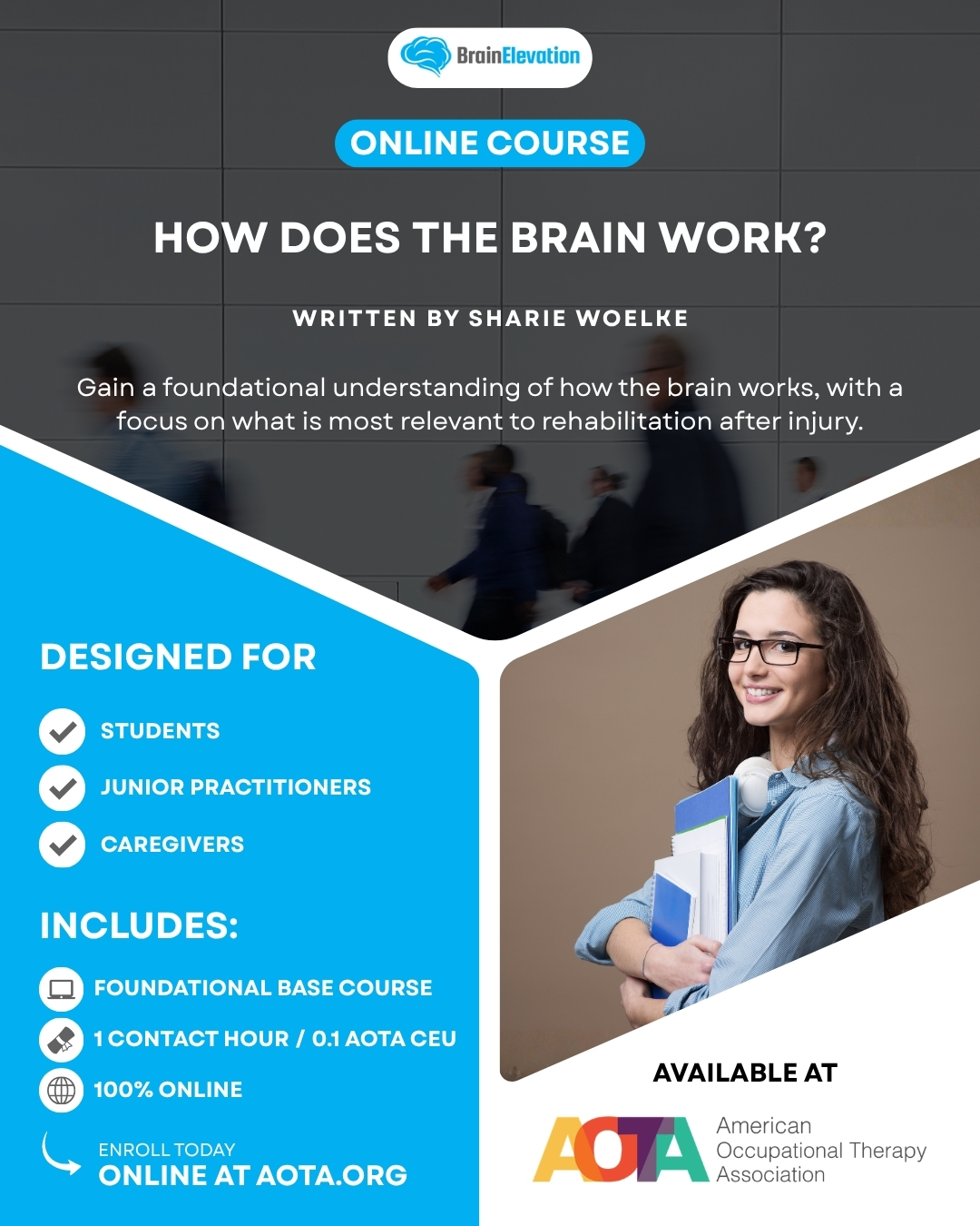
To bridge this exact gap for developing practitioners, Brain Elevation highlights the foundational course: How Does the Brain Work? | Foundations and Early Recovery in Brain Injury Rehabilitation Micro Credential.
Authored by Sharie Woelke, OT Reg. (Ont), RP, BMR, MEd, Clinical Director of Renew Neurotherapy and Director of Instructional Design for Brain Elevation Consulting, this intensive program provides the precise foundational knowledge most relevant to rehabilitation after injury. This course serves as the vital base for the rest of the Professional Certificate in Applied Brain Injury Rehabilitation, seamlessly transitioning your expertise from understanding baseline brain function to actively addressing injury and managing long-term recovery.
Course Specifications:
- Educational Credit: .1 AOTA CEU / 1 contact hour / 1.25 NBCOT PDUs.
- Curriculum Alignment: Part of the Professional Certificate in Applied Brain Injury Rehabilitation and the Brain Basics for Rehabilitation Practice and Recovery Microcredential.
- Core Competencies: Structural protective frameworks, neuronal and glial dynamics, Brodmann area mapping, and Menon’s Triple Network Theory.
Elevate your clinical authority, build a rock-solid scientific foundation, and better serve your brain injury clients by enrolling directly through the American Occupational Therapy Association (AOTA).